Showing posts with label Emil Selenka. Show all posts

Friday, 26 February 2016

Placentation in opossums

Embryo of Virginia Opossum (Didelphys virginiana) at 6 days
of gestation. Opened to show the vessels of the trilaminar yolk sac
and free-floating allantoic sac. From Selenka 1886.
Emil Selenka did not confine himself to rodents and primates (previous post and recent review). He also gave the first detailed description of embryonic development in a marsupial, the Virginia opossum. He bred them in the laboratory and thus had dated pregnancies.

All marsupials have a yolk sac placenta with both two-layered and three-layered areas (bilaminar and trilaminar omphalopleure); the latter has blood vessels that radiate from a sinus terminalis as can be seen in Selenka's illustration.


Neonate of Virginia Opossum. From Selenka 1886.
The Virginia opossum has a pretty short gestation even for a marsupial. The neonate above was born after 13 days. Development is supported largely by histotrophic nutrition, i.e. uptake of uterine gland secretions. The vascular part of the yolk sac is thought to be more important for gaseous exchange.

Note that the allantois makes no contact with the trophoblast. Unlike in the koala, wombat and bandicoots, there is nothing approaching chorioallantoic placentation. The allantois serves mainly as a receptacle for urine excreted by the mesonephros (here). 
Virginia Opossum (D. virginiana) by Cody Pope CC BY-SA 2.5
(Wikipedia Commons)
 
There are 87 species of didelphids in Central and South America, but the Virginia opossum is alone in extending its range to the USA and Canada.

Despite the species richness, placentation has been described for only five opossums; three from the same genus (D. virginiana, D. aurita and D. marsupialis) plus the gray short-tailed opossum (Monodelphis domestica) and gray four-eyed opossum (Philander opossum). I will save them for a later post.

Wednesday, 25 November 2015

Mouse and human blastocysts compared


Mouse blastocyst with trophectoderm (TE),
epiblast (EPI) and primitive endoderm (PE)  from Selenka 1883
Even before implantation, three cell lineages are apparent in the blastocyst of mouse and human. Outermost is the trophectoderm that will contribute trophoblast to the placenta. The inner cell mass has already differentiated into the epiblast and the primitive endoderm or hypoblast. For mouse, this much has been clear since the pioneering studies of Emil Selenka (here).

Gene expression in these three lineages of mouse placenta has been known for some time. What does this tell us about human preimplantation development? A recent paper in Development (here) suggests less than some might like to think.

  • For trophectoderm, key lineages expressed in the mouse (e.g. Id2, Elf5, Eomes) either are not expressed in human trophectoderm or are expressed in alternative lineages.
  • There are several genes that are exclusively expressed in human epiblast (e.g.the transcription factor KLF17).
  • Expression of some genes in primitive endoderm is conserved between mouse and human (e.g. Foxa2/FOXA2).

These findings support other work indicating that the genes and signalling pathways involved in lineage specification differ between mouse and human blastocysts.

Tuesday, 3 November 2015

Placentation in gibbons


Agile Gibbon (Hylobates agilis) Bristol Zoo Gallery
A recent paper on a fossil ape (here) highlights the divergence of the lesser apes (gibbons and siamangs) from the great apes (orangutans, gorilla, chimpanzee, bonobo, human). The fossil (Pliobates cataloniae) has a combination of primitive and derived features that make it difficult to place on the evolutionary tree (discussed here).
 
Gibbons themselves have placentation with some monkey-like features and others shared with the great apes.
 
Uterus of an agile gibbon (H. agilis) opened to show the decidua
capsularis enclosing the embryo. From Selenka 1899
The most important shared characteristic is that the fetus develops beneath a decidua capsularis (see previous post) implying that implantation is interstitial as in great apes. In Old World monkeys implantation is superficial and no decidua capsularis is found. 
 
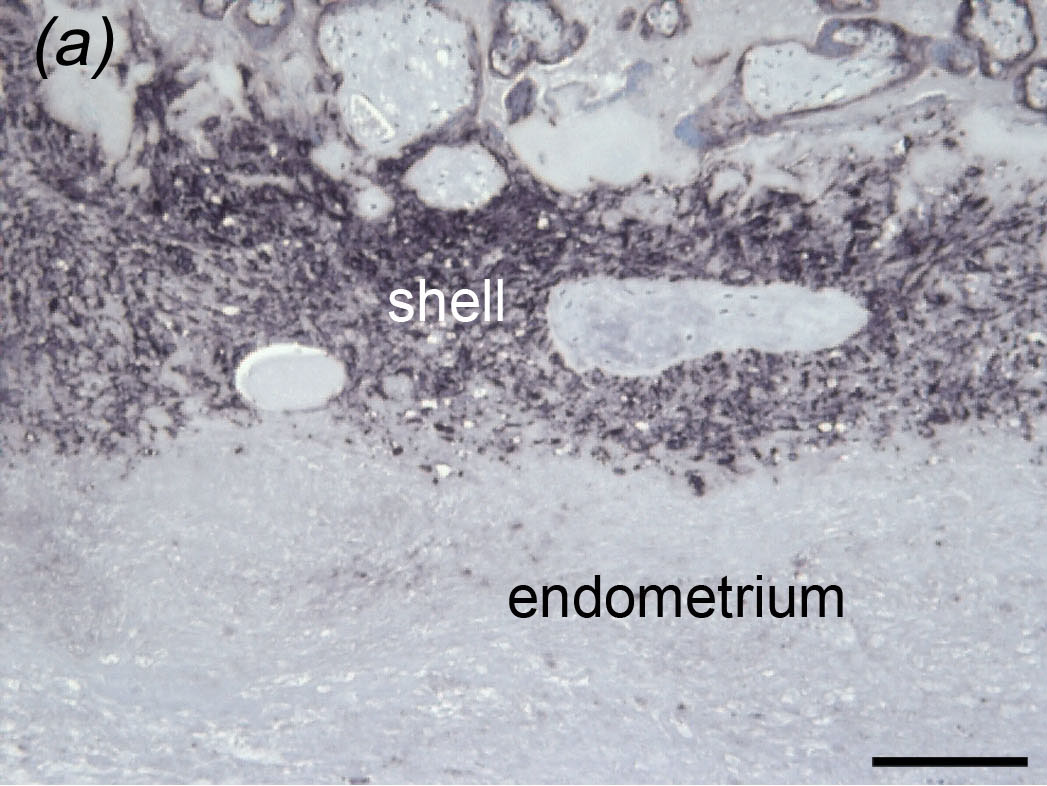
Placental bed of a Javan gibbon (Hylobates moloch)
Reproduced from Carter et al. (c) Museum for Naturkunde Berlin
However, when we examined the placenta of a Javan gibbon we found a continuous trophoblastic shell and a sharp boundary between the shell and the underlying endometrium - just as in Òld World monkeys. In great apes, the boundary is less distinct because trophoblast cells invade the endometrium by this route.

Wednesday, 18 February 2015

Selenka's gibbons

Bornean White-bearded Gibbon (Hylobates albibarbis)
Primate Info Net (University of Wisconsin) Photo Credit Marilyn Cole
Emil Selenka showed that the gibbon embryo, like that of humans and other apes, develops in the uterine wall beneath a decidua capsularis. But what species did he study?

Most of his figures are of a gibbon identified as Hylobates concolor (Harlan) from Borneo. The species name is still in use for Nomascus concolor, which is not found on Borneo. I now know, thanks to Dr. Thomas Geissman and his remarkable web site, that this reflects an extraordinary comedy of errors. Harlan described his ape as a hermaphrodite orangutan from Borneo; in fact it was a juvenile gibbon from Indochina!

Geographical distribution of gibbons.
(C) 2010 Thinh et al.
How then can we identify Selenka's gibbon? A study of mitochondrial genes (here) concluded that there were two species of gibbon on Borneo, one of them with three subspecies. Fortunately Selenka stated his specimens were collected on the left bank of the Kapuas River, in the territory occupied by the Bornean White-bearded Gibbon (Hylobates albibarbis) shown above.

Early stage of pregnancy in Hylobates albibarbis with amnion (A), yolk sac (D) and
exocoelom (Ex). The specimen had been flattened by contraction of the uterus but
the decidua capsularis (Dc) is clearly seen.
The embryo is depicted above. It had a primitive streak but no somites. Therefore it may correspond to Carnegie Stage 7 or early Stage 8 in the human. Selenka's paper can be found on the web (read only).

Wednesday, 21 January 2015

Emil Selenka

Emil Selenka (27 February 1842 - 21 January 1902
Emil Selenka died 113 years ago today, just before his 60th birthday. He was an eminent German zoologist, who spent much of his career studying the development of marine invertebrates, especially sea cucumbers. He turned to vertebrates rather late but made important contributions to the embryology and placentation of primates. His work on gibbons and orangutans is especially significant.

Pregnant uterus of Hylobates agilis (rafflei) showing the decidua
capsularis (d.c.) reproduced by Hill (here) from Selenka

I have shown this image before to document that gibbons resemble other great apes in having a decidua capsularis. Thus implantation is interstitial as it is in the human.

Fetus of Hylobates muelleri (Müller's Bornean Gibbon) and
uterus of Nomascus concolor (Black Crested Gibbon) from Selenka
In a moving tribute to Selenka, his pupil Hubrecht (previous post) wrote that his artistic talent was so great that it was almost a pity he became a professor of zoology rather than a painter.

Selenka amassed a large number of specimens and kept the skeletons as well as the reproductive tracts. He felt the maximum information should be gained from the animals that lost their lives. One recent study (here) estimates that Hubrecht may have bagged as many as 400 orangutans between 1892 and 1895.