Friday, 13 November 2015

The mouse yolk sac

Placenta Volume 36 Issue 10 October 2015
Great to see yolk sacs on the front cover of Placenta. Too often the mouse yolk sac is discarded or ignored in the laboratory - either through ignorance or because it has no obvious equivalent in human pregnancy. This particular study showed how mouse deficient in the OGA gene have defective blood vessel formation (vasculogenesis). This affected branching of the blood vessels in the yolk sac.

Ignorance of the yolk sac is evidenced by a high impact journal publishing a schematic where mouse yolk sac is shown to be only residual as occurs in humans (here).

I believe the mouse model of placentation is incomplete unless the yolk sac is studied along with the chorioallantoic placenta (argued here).

Friday, 6 November 2015

Placentation in the ferret

Transverse section through the uterus of a ferret (Mustela putorius furo)
at 28 days of gestation. From Strahl and Ballmann 1915.
A, allantois; H, hemophagous organ; N. yolk sac
A recent paper discussed the domestic ferret as a model for perinatal brain injury. As  the ferret is an altricial species, events that occur during human pregnancy are deferred to the postnatal period. However, neurogenesis and neuronal migration start in pregnancy and the effects of hypoxia on these processes could be explored.

Electron micrograph of the interhemal region in a ferret placenta.
Courtest of Dr. Allen C. Enders
Like all carnivores (except hyenas), ferrets have an endotheliochorial placenta. The endothelium of the maternal capillaries is swollen as readily seen by comparing with the endothelium of fetal capillaries above.

The interhemal region of the ferret placenta.
Courtesy of Dr. Allen C. Enders
At low power it can be seen that the fetal capillaries indent the surrounding trophoblast so the diffusion distance from maternal to fetal blood is minimized. Thus oxygen should diffuse across this barrier almost as readily as in the human hemochorial placenta.

Hematoidin crystals in the hemophagous region of the ferret placenta.
From Strahl and Ballmann 1915
Like other carnivores, the ferret has a prominent hemophagous region (shown in the top figure). Here trophoblast takes up maternal red blood cells by phagocytosis and processes the contained hemoglobin to extract the iron. The hemoglobin breakdown product is hematoidin, which crystallizes out. We have described the same thing in tenrec placenta (here).

The most thorough study of ferret placenta was published a century ago (full reference here). The ultrastructure was later described by Lawn and Chiquoine (here). There are recent reviews of endotheliochorial placentation (here) and placentation in carnivores (here).

Tuesday, 3 November 2015

Placentation in gibbons


Agile Gibbon (Hylobates agilis) Bristol Zoo Gallery
A recent paper on a fossil ape (here) highlights the divergence of the lesser apes (gibbons and siamangs) from the great apes (orangutans, gorilla, chimpanzee, bonobo, human). The fossil (Pliobates cataloniae) has a combination of primitive and derived features that make it difficult to place on the evolutionary tree (discussed here).
 
Gibbons themselves have placentation with some monkey-like features and others shared with the great apes.
 
Uterus of an agile gibbon (H. agilis) opened to show the decidua
capsularis enclosing the embryo. From Selenka 1899
The most important shared characteristic is that the fetus develops beneath a decidua capsularis (see previous post) implying that implantation is interstitial as in great apes. In Old World monkeys implantation is superficial and no decidua capsularis is found. 
 
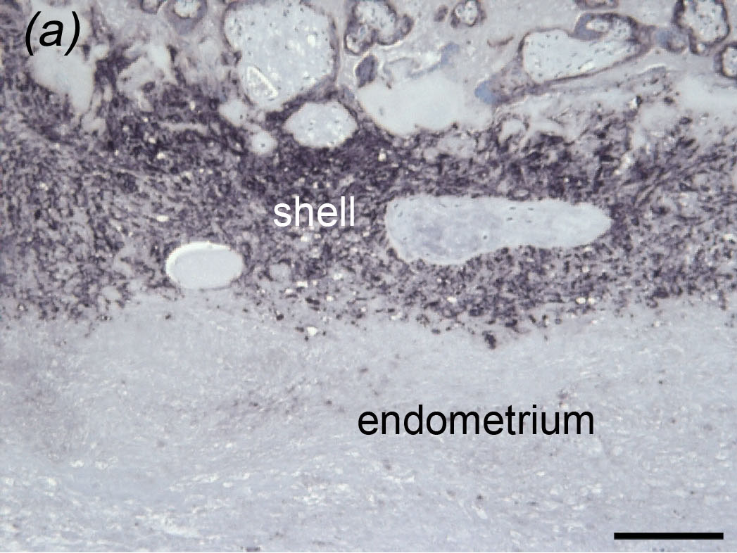
Placental bed of a Javan gibbon (Hylobates moloch)
Reproduced from Carter et al. (c) Museum for Naturkunde Berlin
However, when we examined the placenta of a Javan gibbon we found a continuous trophoblastic shell and a sharp boundary between the shell and the underlying endometrium - just as in Òld World monkeys. In great apes, the boundary is less distinct because trophoblast cells invade the endometrium by this route.

Friday, 23 October 2015

Clan of the cave bear: researching ancient DNA

Reconstruction of a cave bear (Ursus spelaeus)
Uploaded by Sergiodlarosa to Wikimedia Commons (CC)
The proceedings of a Royal Society discussion meeting on ancient DNA have just been published (here). Three reviews in particular captured my interest.

Ancient DNA: the first three decades by Hagelberg, Hofreiter and Keyser (here) is a lucid account of the history of the field (a major advance was shotgun sequencing as applied to the cave bear). It highlights not only high profile papers from Nature and Science (some of them reviewed in my blog), but also gives credit to important follow up studies from specialist journals. A very useful ressource indeed!


Ancient genomics by Der Sarkissian et al. (here) is from the renowned Centre for GeoGenetics in Copenhagen. This review is especially strong on the technical advances in the field and even includes a user's manual. With the techniques initially available it would have required 180 kg of material and 130 million amplicons to generate a first draft of the cave bear genome. A lot has happened since then. They conclude that even "Looking back 5 years, no one could have predicted the current state of current genomics."

Almost 20 years of Neanderthal palaeogenetics by Sánchez-Quinto and Lalueza-Fox (here) does a remarkably good job of surveying what has been learned from the DNA of Neanderthals from various geographical locations. It covers more than just their relations to and interbreeding with Denisovans and modern humans (previous post). A great deal can be inferred about their demographics, population size and ultimate extinction.

ISBN 0-517-54202-1

 The title of this post is of course an homage to the fiction of Jean M. Auel. The clan of the cave bear are Neanderthals who interact (and interbreed) with modern humans. First published in 1980 it far anticipated the scientific evidence given in the above reviews.

Wednesday, 14 October 2015

Pregnancy-associated malaria research suggests a way to target cancer cells

Anopheles stephensi - a vector for the malaria parasite
Centers for Disease Control and Prevention

The malaria parasite Plasmodium falciparum is spread by mosquitos. It multiplies and resides in the liver and erythrocytes (red blood cells). In areas with heavy infection with Plasmodium most individuals acquire immunity. During pregnancy, however, infected erythrocytes are able to accumulate in the placenta. Pregnancy-associated malaria is an important cause of maternal and fetal morbidity and mortality (reviewed here).


Placenta from a stillbirth due to maternal malaria infection. Normal erythrocytes lack
a nucleus. Thus a nucleus indicates the presence of the malaria parasite.
Wikimedia Commons (CC BY-SA 3.0) uploaded by Nephron
Infected erythrocytes express surface proteins, among them VAR2CSA, which can bind to a placental variant of chondroitin sulphate A (CSA) expressed by syncytiotrophoblast.

A paper just out in Cancer Cell (here) shows that placental CSA is expressed by many types of cancer cell. This knowledge was leveraged to target cancer cells using recombinant VAR2CSA fused to diptheria toxin or conjugated to hemiasterlin. Human cancer cells in vitro were effectively killed by this means. Tumour growth and metastasis in mice could be inhibited by a similar approach.

Sunday, 11 October 2015

A. C. Haddon

Mask of turtle shell plates made by Torres Strait Islander and described by A. C. Haddon
Creative Commons (CC BY-NC-SA 4.0) The Trustees of the British Museum
Alfred Cort Haddon was a marine zoologist who  morphed into an eminent ethnologist and anthropologist after joining an expedition to the Torres Strait in 1898.

The Torres Strait Islanders are a Melanesian people distinct from the Aborigines of mainland Australia. Haddon collected their artefacts avidly -- convinced that their culture would soon be repressed by zealous missionaries. I had an opportunity to view some of these artefacts on a recent visit to the Queensland Museum in Brisbane. Haddon donated also to the British Museum, but the bulk of his Collection is now in the Museum of Anthropology and Archaeology at Cambridge University.

Alfred Cort Haddon (1855-1940)
The Wellcome Museum, Wellcome Images (CC BY 4.0)
Haddon's Collection was key to a 2011 study that examined the origin of the Australian Aborigines (here). A genomic sequence was obtained from a hair sample that had been collected in the 1920s as Haddon passed through Golden Ridge, near Kalgoorlie, Western Australia. Ethical concerns about using this material were allayed when it could be shown, with the aid of contemporary newspaper reports, that the sample was donated voluntarily. The main finding was that Aborigines are descendents of a human dispersal out of Africa that was separate and much earlier than that giving rise to present day Asians.


Cambridge University Press 1924 (Second and Revised Edition)
Human genomics has greatly improved our understanding of the migrations that gave rise to modern peoples. Haddon would have been fascinated. He did what he could with the tools then available to him and summarized them in the above book. The title was not as controversial at the time as it might be considered today.

Friday, 2 October 2015

Transcriptome of the pregnant male seahorse

Pot-bellied seahorse (Hippocampus abdominalis) from Sketchbook of Fishes
by William Buelow Gould (1801-53)
Viviparity in seahorses involves incubation of the embryos in the brood pouch of the male. A new study (abstract here) analyses the transcriptome of the brood pouch of the pot-bellied seahorse for genes that are upregulated during pregnancy and in transition to the post partum state.

Many of the same genes or their homologs are upregulated during pregnancy in mammals and other viviparous taxa. Placentologists will want to scan the tables for their favourite genes. I found it interesting that genes involved in lipid transport and iron transfer are upregulated.

The authors suggest the possibility that a common toolkit of genes is recruited to support pregnacy in mammals, reptiles and live-bearing fish.