Tuesday, 12 January 2016

Steller's sea cow

Reconstruction of Steller's sea cow (Hydrodamalis gigas)
By Emőke Dénes (Natural History Museum in London) (CC BY-SA 2.5)
When first described by Steller in 1741, the range of this sirenian was already restricted. Intensification of hunting led to extinction of the species within another 27 years.

A recent paper with a "stellar" cast of authors (here) uses molecular data to firmly establish the position of Steller's sea cow within crown Afrotheria; with additional morphological data it also shows shows how the species fits into the fossil record.

Steller's sea cow (Hydrodamalis gigas) belongs with the dugong (Dugong dugon) in Family Dugongidae whereas the three species of manatee belong to Trichechidae. Together they make up the Order Sirenia, which is sister to the elephant Order Proboscidea.

Endotheliochorial placenta of the Amazonian manatee
Fetal (fc) and maternal (mc) capillaries are present
Elephants have an endotheliochorial placenta. Therefore it perplexed us that Wislocki had described the placenta of the West Indian manatee (Trichechus manatus) as haemochorial. Therefore we reopened the matter by studying placentas from the Amazonian manatee (T. inunguis). We were able to demonstrate two sets of capillaries (shown above) and conclude this placenta to be endotheliochorial (here).

The allantoic sac of elephants and manatees has four chambers and we have shown this to be a shared, derived character of the superorder Afrotheria (here).

Wednesday, 6 January 2016

Placentation in salps

Placenta of a salp (Salpa fusiformis)
Reproduced from Bone, Pulsford and Amoroso (here)
(C) 1985 with permission from Elsevier
Sea squirts? asked a faithful reader of this blog. OK, sea squirts do not have placentas, but salps do! They, too, are tunicates, and thus included in the sister group to vertebrates (previous post). Whilst sea squirts are mainly sessile, salps are pelagic, living near the surface of oceans and at times more numerous than krill. They are tubular animals and swim by jet propulsion.

The life cycle involves alternation between asexual and sexual generations. The asexual phase (oozooid) develops within the jet chamber of the sexual phase (blastozooid). The image above is from Amoroso's last paper, published almost three years after his death (see previous post for Amo). The placenta consists of two layers: an outer cortex (co) and an inner central layer (c). These separate the embryonic (E) and maternal (M). circulations. Both layers are syncytial and both are maternal in origin. However, embryonic leucocytes pass into and add to the cortical layer.

Blastozooids are hermaphrodite, but the egg develops before the testis matures so is fertilized by sperm from a different blastozooid

HMS Rattlesnake on which Thomas Henry Huxley served as
Assistant Surgeon during the voyage to Australia and New Guinea 1846-50
National Maritime Museum (public domain)
The first English language description of placentation in the Family Salpida was given by T. H. Huxley R.N., "late of HMS Rattlesnake," in 1851 (here) although he cites even earlier work by Cuvier, Chamisso and Meyen.

We tend to think of placentation in terms of mammals, reptiles and fish, but a current paper in Biological Reviews (here) shows that maternal provision of nutrients (matrotrophy) and even placentation is not infrequent in invertebrate phyla.

Monday, 30 November 2015

Sea squirts, lancelets and acorn worms

A sea squirt (Ciona intestinalis) - a member of Tunicata 
Wikimedia Commons uploaded by perezoso (GFDL)
Genomics has clarified our position in the tree of life. To explain this I need to define some taxonomic terms.

Phylum Chordata comprises three subphyla: Vertebrata (Craniata), Tunicata (Urochordata)and Cephalochordata. Tunicates include sea squirts  such as Ciona (pictured) above. A familiar cephalochordate is the lancelet Branchiostoma lanceolatum better known as Amphioxus (shown below).

Amphioxus or Branchiostoma lanceolatum
(c) Virginia Gewin here (CC-BY-SA 3.0)
Amphioxus has long been used to exemplify the general plan of chordate organization and lancelets used to be regarded as the closest relatives to vertebrates. The genomic evidence, however, has tunicates like the sea squirts as sister group to vertebrates with cephalochordates as a deeper branch. Additional support is given by conserved molecular signatures (here).

Chordates belong in the Superphylum Deuterostomia (brilliantly reviewed by Lowe et al. here) along with Phylum Hemichordata and Phylum Echinodermata. Echinoderms are richly represented in the fossil record and the five extant classes include sea urchins, sea cucumbers and starfish. Hemichordates include the acorn worms for which two genomes just became available (here and here).

Acorn Worms (Hemichordata: Enteropneusta)
from Spengel 1883 (public domain)
One of many findings was a cluster of six genes that are conserved across chordates and implicated in patterning of gill slits. This is significant because gill slits were an innovation in the deuterostome lineage (although secondarily lost in echinoderms).

Relationships between deuterostome phyla were largely worked out through their embryology, an example being the erection of Chordata by Haeckel. Understanding the genes involved in developmental processes remains a focus in working out our evolutionary history (see the review by Lowe et al. mentioned above).

Wednesday, 25 November 2015

Mouse and human blastocysts compared


Mouse blastocyst with trophectoderm (TE),
epiblast (EPI) and primitive endoderm (PE)  from Selenka 1883
Even before implantation, three cell lineages are apparent in the blastocyst of mouse and human. Outermost is the trophectoderm that will contribute trophoblast to the placenta. The inner cell mass has already differentiated into the epiblast and the primitive endoderm or hypoblast. For mouse, this much has been clear since the pioneering studies of Emil Selenka (here).

Gene expression in these three lineages of mouse placenta has been known for some time. What does this tell us about human preimplantation development? A recent paper in Development (here) suggests less than some might like to think.

  • For trophectoderm, key lineages expressed in the mouse (e.g. Id2, Elf5, Eomes) either are not expressed in human trophectoderm or are expressed in alternative lineages.
  • There are several genes that are exclusively expressed in human epiblast (e.g.the transcription factor KLF17).
  • Expression of some genes in primitive endoderm is conserved between mouse and human (e.g. Foxa2/FOXA2).

These findings support other work indicating that the genes and signalling pathways involved in lineage specification differ between mouse and human blastocysts.

Friday, 13 November 2015

The mouse yolk sac

Placenta Volume 36 Issue 10 October 2015
Great to see yolk sacs on the front cover of Placenta. Too often the mouse yolk sac is discarded or ignored in the laboratory - either through ignorance or because it has no obvious equivalent in human pregnancy. This particular study showed how mouse deficient in the OGA gene have defective blood vessel formation (vasculogenesis). This affected branching of the blood vessels in the yolk sac.

Ignorance of the yolk sac is evidenced by a high impact journal publishing a schematic where mouse yolk sac is shown to be only residual as occurs in humans (here).

I believe the mouse model of placentation is incomplete unless the yolk sac is studied along with the chorioallantoic placenta (argued here).

Friday, 6 November 2015

Placentation in the ferret

Transverse section through the uterus of a ferret (Mustela putorius furo)
at 28 days of gestation. From Strahl and Ballmann 1915.
A, allantois; H, hemophagous organ; N. yolk sac
A recent paper discussed the domestic ferret as a model for perinatal brain injury. As  the ferret is an altricial species, events that occur during human pregnancy are deferred to the postnatal period. However, neurogenesis and neuronal migration start in pregnancy and the effects of hypoxia on these processes could be explored.

Electron micrograph of the interhemal region in a ferret placenta.
Courtest of Dr. Allen C. Enders
Like all carnivores (except hyenas), ferrets have an endotheliochorial placenta. The endothelium of the maternal capillaries is swollen as readily seen by comparing with the endothelium of fetal capillaries above.

The interhemal region of the ferret placenta.
Courtesy of Dr. Allen C. Enders
At low power it can be seen that the fetal capillaries indent the surrounding trophoblast so the diffusion distance from maternal to fetal blood is minimized. Thus oxygen should diffuse across this barrier almost as readily as in the human hemochorial placenta.

Hematoidin crystals in the hemophagous region of the ferret placenta.
From Strahl and Ballmann 1915
Like other carnivores, the ferret has a prominent hemophagous region (shown in the top figure). Here trophoblast takes up maternal red blood cells by phagocytosis and processes the contained hemoglobin to extract the iron. The hemoglobin breakdown product is hematoidin, which crystallizes out. We have described the same thing in tenrec placenta (here).

The most thorough study of ferret placenta was published a century ago (full reference here). The ultrastructure was later described by Lawn and Chiquoine (here). There are recent reviews of endotheliochorial placentation (here) and placentation in carnivores (here).

Tuesday, 3 November 2015

Placentation in gibbons


Agile Gibbon (Hylobates agilis) Bristol Zoo Gallery
A recent paper on a fossil ape (here) highlights the divergence of the lesser apes (gibbons and siamangs) from the great apes (orangutans, gorilla, chimpanzee, bonobo, human). The fossil (Pliobates cataloniae) has a combination of primitive and derived features that make it difficult to place on the evolutionary tree (discussed here).
 
Gibbons themselves have placentation with some monkey-like features and others shared with the great apes.
 
Uterus of an agile gibbon (H. agilis) opened to show the decidua
capsularis enclosing the embryo. From Selenka 1899
The most important shared characteristic is that the fetus develops beneath a decidua capsularis (see previous post) implying that implantation is interstitial as in great apes. In Old World monkeys implantation is superficial and no decidua capsularis is found. 
 
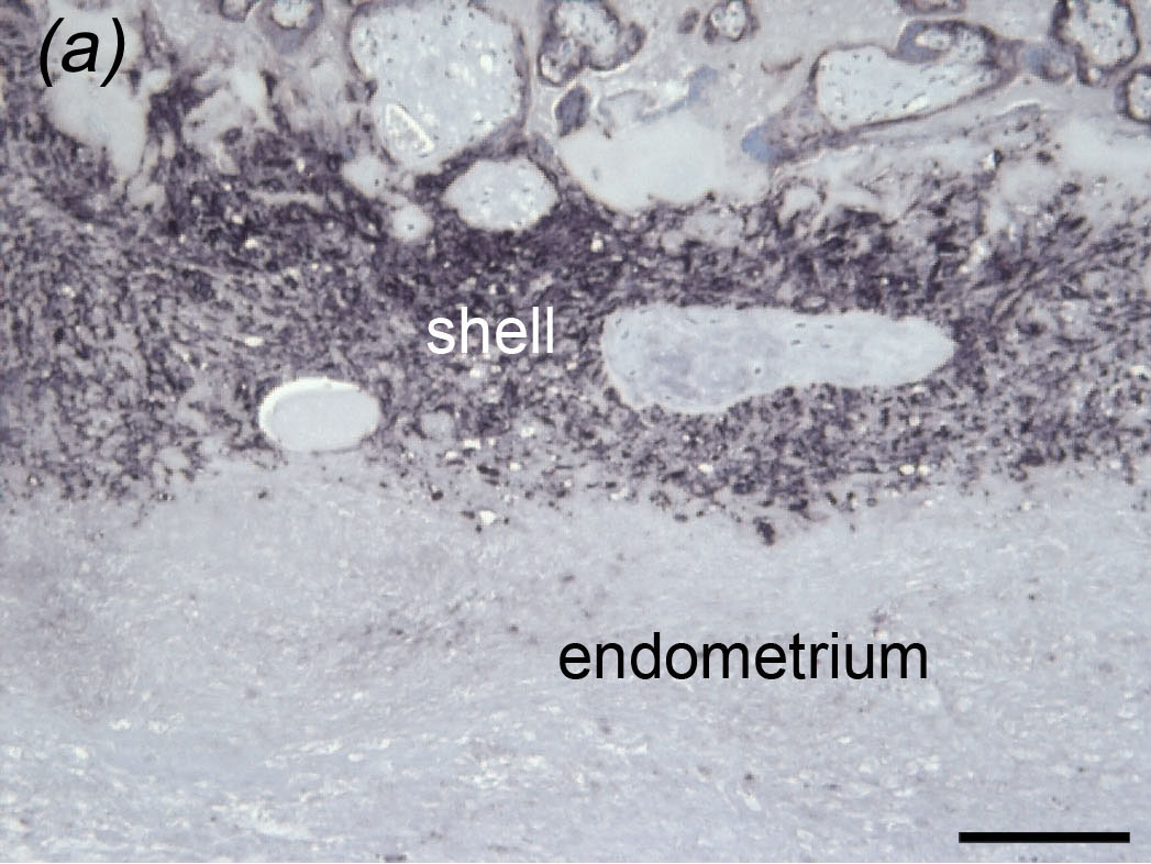
Placental bed of a Javan gibbon (Hylobates moloch)
Reproduced from Carter et al. (c) Museum for Naturkunde Berlin
However, when we examined the placenta of a Javan gibbon we found a continuous trophoblastic shell and a sharp boundary between the shell and the underlying endometrium - just as in Òld World monkeys. In great apes, the boundary is less distinct because trophoblast cells invade the endometrium by this route.